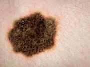
Calculated tumor area is a novel, microscopic feature and a two-dimensional surrogate
for tumor size
MONDAY, July 1, 2019 (HealthDay News) — Stratification of melanoma into groups based on calculated tumor area improves prognostic value over stratification using the category based on Breslow thickness, according to a study published online June 26 in JAMA Dermatology.
Gerald Saldanha, F.R.C.Path., from the University of Leicester in the United Kingdom, and colleagues developed a two-dimensional feature, calculated tumor area (CTA). The Leicester development sample included 918 patients with available primary tumor tissue, a diagnosis from 2004 through 2011, and invasive disease. The Nottingham validation sample included 321 patients with an anonymized spreadsheet with primary melanoma diagnosed from 2003 through 2005, or from 2008 through 2010.
The researchers found that in the Leicester cohort, CTA was an independent prognostic factor after adjusting for Breslow thickness, age, sex, ulcer, mitotic rate, and microsatellites (hazard ratio [HR], 1.87). In the validation cohort, the CTA had an HR of 1.55. When CTA was not in the model, Breslow thickness was significant in multivariable analysis only. However, the relative importance of CTA was shown by its retention in all 100 bootstrap multivariable models with backward selection versus 53 that retained Breslow thickness. CTA stratification of melanomas showed wider separation of survival curves compared to those stratified using Breslow thickness (HRs, 1.00 to 41.46 and 1.00 to 36.95, respectively).
“These findings suggest that calculated tumor area should be prioritized for investigation to verify its prognostic value and to assess its applicability and acceptability in routine practice,” the authors write.
Editorial (subscription or payment may be required)
Copyright © 2019 HealthDay. All rights reserved.


